Moody JB, Poitrasson-Rivière A, Renaud JM, Hagio T, Alahdab F, Al-Mallah MH, Vanderver MD, Goonewardena SN, Ficaro EP, Murthy VL. (2025). A foundation transformer model with self-supervised learning for ECG-based assessment of cardiac and coronary function.
Description:
A new study in NEJM AI describes a transformer-based electrocardiogram (ECG) “foundation” model developed by researchers at INVIA Medical Imaging Solutions and partners. Instead of relying on massive labeled datasets, which are difficult and expensive to create, this model learned from more than 800,000 unlabeled ECGs, then fine-tuned with smaller sets of high-quality labels from PET, SPECT, MRI, and clinical ECG reports.
The result is a single, versatile model capable of performing cardiac prediction tasks, ranging from myocardial flow reserve (MFR) to LVEF and automated ECG interpretation. It captures subtle physiologic patterns in the ECG that traditional models often miss, especially in areas like ischemia and coronary microvascular dysfunction, where labeled training data are limited.
Key Findings:
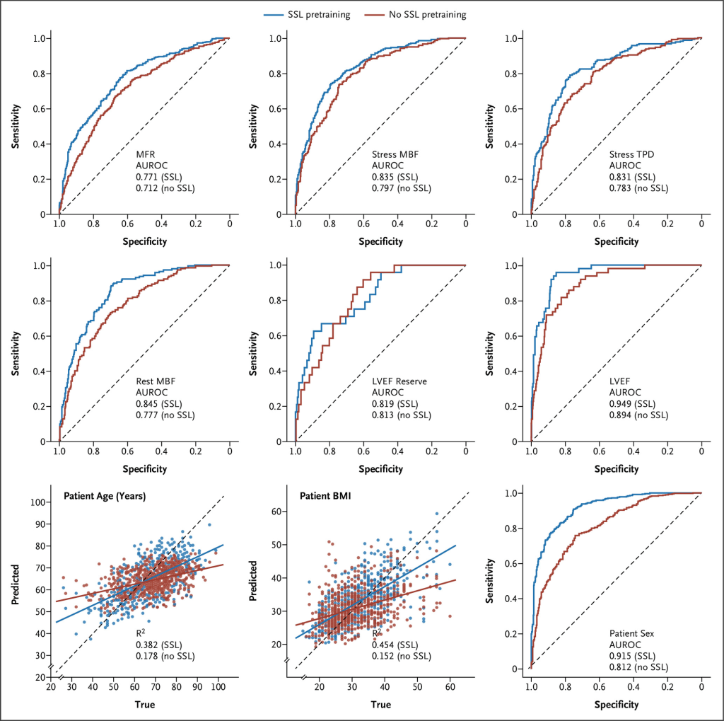
Across five independent testing cohorts, the SSL-pretrained model consistently outperformed the same architecture trained from scratch. In fact, it improved diagnostic accuracy for 11 out of 12 tasks. For example, in an external PET cohort, performance for detecting impaired MFR rose from 0.712 to 0.771 AUROC, and accuracy for identifying impaired LVEF increased from 0.894 to 0.949 with SSL pretraining.
What’s equally impressive is how efficiently the model learns. For some tasks, like predicting LVEF, it reached the same accuracy as supervised training while using less than 20% of the labeled data. And even when tested on completely different imaging modalities, such as SPECT or MRI, the model continued to perform strongly, demonstrating that the foundation approach generalizes well across patient populations and clinical environments.
Clinical Relevance:
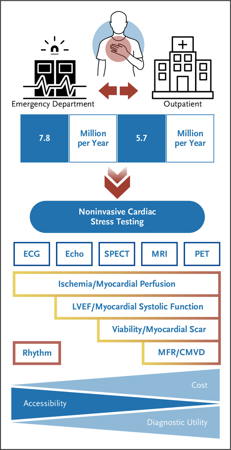
This work has meaningful implications for how clinicians may assess cardiac and coronary health in the future. Key physiologic markers like myocardial flow reserve, one of the strongest indicators of coronary microvascular dysfunction—are typically only available through advanced imaging like PET. Because PET is expensive and not widely accessible, many patients who could benefit from physiologic assessment never receive it.
By showing that MFR, stress MBF, and other advanced metrics can be estimated from a standard 10-second ECG, this research opens the door to more accessible and cost-effective evaluation tools. An ECG-based model could help clinicians identify patients who need further testing, enhance the value of existing stress tests, and ultimately make it easier to detect conditions like microvascular disease earlier in the care pathway.
For INVIA, this study also reinforces the importance of high-quality quantitative imaging. PET and SPECT datasets processed with software such as 4DM provide precise perfusion and function measurements that can be used to supervise advanced ECG models.
Partners in Research:
INVIA Medical Imaging Solutions, the Division of Cardiovascular Medicine in the Department of Internal Medicine at the University of Michigan, the Department of Cardiology at the University of Missouri, the Houston Methodist DeBakey Heart and Vascular Center and the VA Ann Arbor Healthcare System collaborated on this research.