Moody, J.B., Poitrasson-Rivière, A., Hagio, T. et al. Added value of myocardial blood flow using 18F-flurpiridaz PET to diagnose coronary artery disease: The flurpiridaz 301 trial. J. Nucl. Cardiol. 28, 2313–2329 (2021).
Description:
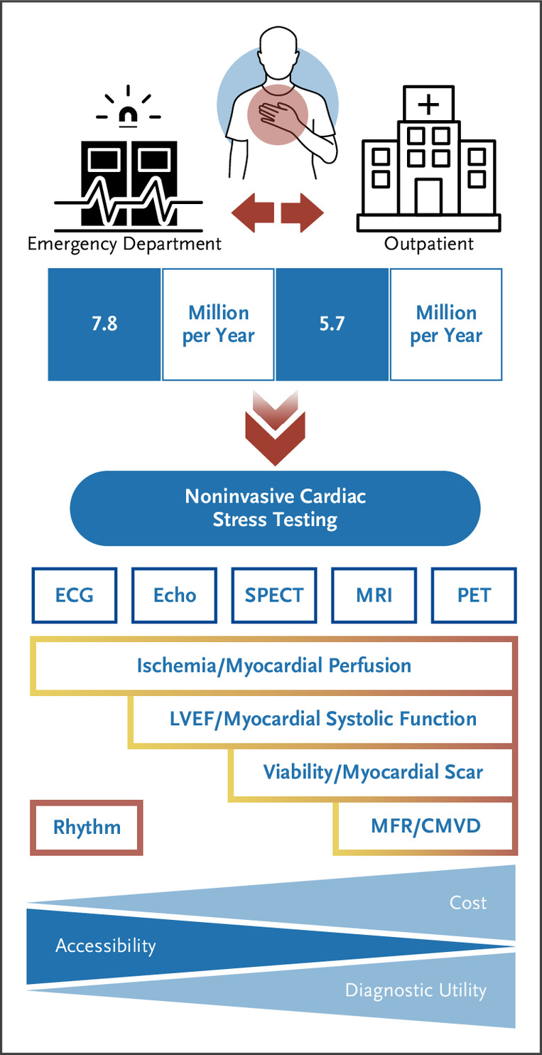
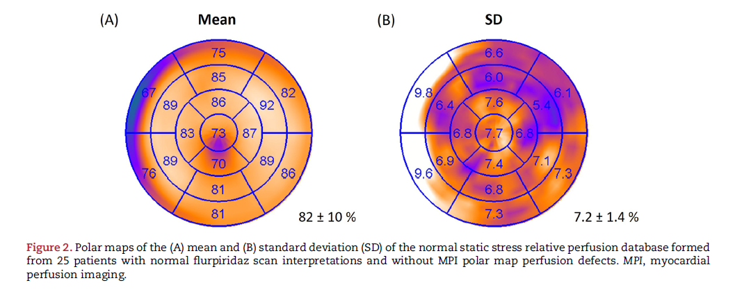
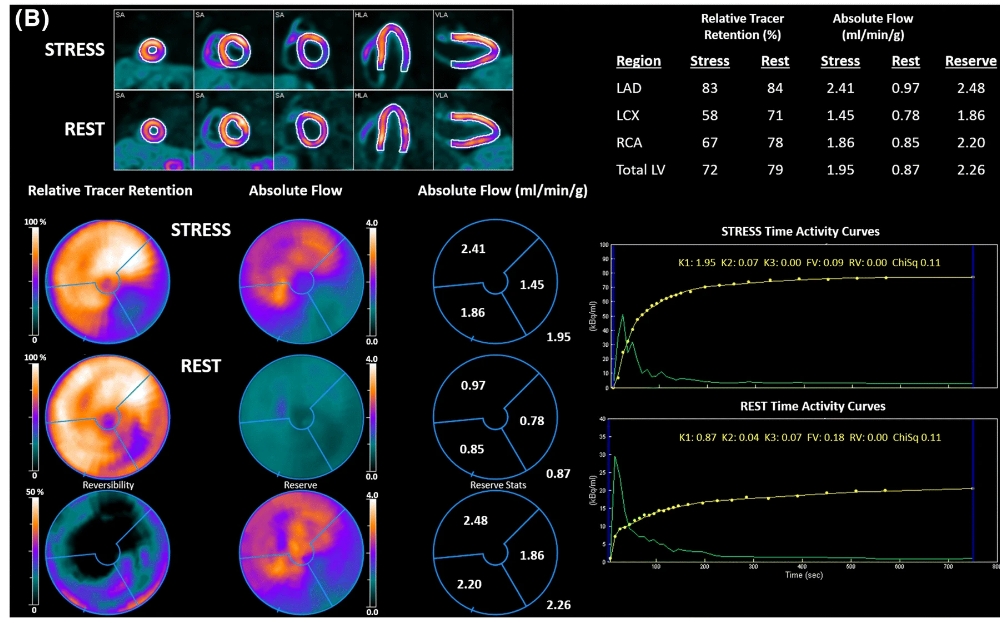
This study validated the incremental diagnostic value of absolute myocardial blood flow (MBF) quantification using the investigational radiotracer 18F-flurpiridaz in a large multicenter trial against quantitative coronary angiography. A subset of 231 patients from the phase 3 flurpiridaz trial was retrospectively analyzed. Dynamic PET data at rest and during pharmacologic stress were used to fit a 2-tissue-compartment model to estimate MBF and myocardial flow reserve (MFR).
The study found that stress MBF and MFR significantly declined with increasing stenosis severity and provided incremental diagnostic value beyond patient characteristics and relative perfusion analysis.
Clinical Relevance:
The findings demonstrate that MBF and MFR quantification using 18F-flurpiridaz PET imaging can accurately identify obstructive coronary artery disease (CAD) and enhance diagnostic precision.
This validation supports the routine clinical application of 18F-flurpiridaz PET for myocardial perfusion imaging, offering a robust tool for improving CAD diagnosis and potentially guiding better-informed treatment decisions. Absolute quantification of myocardial blood flow enhances the ability to assess disease severity, which is crucial for effective risk stratification and management in CAD patients.

Partners in Research:
INVIA Medical Imaging Solutions, GE Healthcare, and the University of Michigan collaborated on this research.